• Baptist Eye Center
    501-221-0123
  • 4261 Stockton Drive
    501-221-0123

Thyroid Gland Anatomy

  • The thyroid is one of the largest endocrine glands in the body. This gland is found in the neck inferior to (below) the thyroid cartilage (may also be called the Adam's apple in men) and at approximately the same level as the cricoid cartilage. The thyroid controls how quickly the body burns energy, makes proteins, and controls how sensitive the body should be to other hormones.
  • The thyroid participates in these processes by producing thyroid hormones, principally thyroxine (T4) and triiodothyronine (T3). These hormones regulate the rate of metabolism and affect the growth and rate of function of several other systems in the body. Iodine and tyrosine are used to form both T3 and T4. The thyroid also produces the hormone calcitonin, which plays a role in calcium homeostasis.
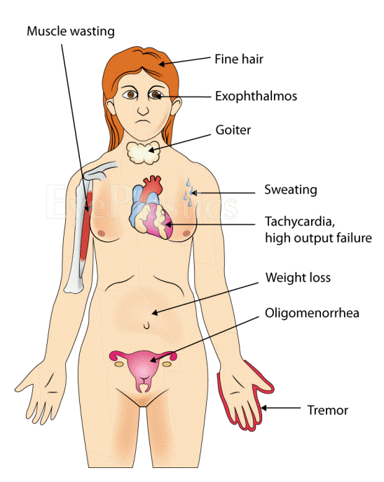
  • The thyroid is controlled by the hypothalamus and pituitary. The gland gets its name from the Greek word for "shield", after the shape of the related thyroid cartilage. Hyperthyroidism (overactive thyroid) and hypothyroidism (underactive thyroid) are the most common problems of the thyroid gland.

Illustration depicting an overview of the thyroid gland and its surrounding structures.

  • The thyroid gland is a butterfly-shaped organ and is composed of two cone-like lobes or wings: lobus dexter (right lobe) and lobus sinister (left lobe), connected with the isthmus. The organ is situated on the anterior side of the neck, lying against and around the larynx and trachea, reaching posteriorly the oesophagus and carotid sheath. It starts cranially at the oblique line on the thyroid cartilage (just below the laryngeal prominence or Adam's apple) and extends inferiorly to the fourth or fifth tracheal ring. It is difficult to demarcate the gland's upper and lower border with vertebral levels because it moves position in relation to these during swallowing.
  • The thyroid gland is covered by a fibrous sheath, the capsula glandulae thyroidea, composed of an internal and external layer. The external layer is anteriorly continuous with the lamina pretrachealis fasciae cervicalis and posteriorolaterally continuous with the carotid sheath. The gland is covered anteriorly with infrahyoid muscles and laterally with the sternocleidomastoid muscle. Posteriorly, the gland is fixed to the cricoid and tracheal cartilage and cricopharyngeus muscle by a thickening of the fascia to form the posterior suspensory ligament of Berry[In variable extent, Lalouette's Pyramid, a pyramidal extension of the thyroid lobe, is present at the most anterior side of the lobe. In this region the recurrent laryngeal nerve and the inferior thyroid artery pass next to or in the ligament and tubercle. Between the two layers of the capsule and on the posterior side of the lobes there are on each side two parathyroid glands.
  • The thyroid isthmus is variable in presence and size, and can encompass a cranially extending pyramid lobe (lobus pyramidalis or processus pyramidalis), remnant of the thyroglossal duct. The thyroid is one of the larger endocrine glands, weighing 2-3 grams in neonates and 18-60 grams in adults, and is increased in pregnancy.
  • The thyroid is supplied with arterial blood from the superior thyroid artery, a branch of the external carotid artery, and the inferior thyroid artery, a branch of the thyrocervical trunk, and frequently by the thyroid ima artery, branching directly from the aortic arch. The venous blood is drained via superior thyroid veins, draining in the internal jugular vein, and via inferior thyroid veins, draining via the plexus thyroideus impar in the left brachiocephalic vein. Lymphatic drainage passes commonly the lateral deep cervical lymph nodes and the pre- and parathracheal lymph nodes. The gland is supplied by sympathetic nerve input from the superior cervical ganglion and the cervicothoracic ganglion of the sympathetic trunk and by parasympathetic nerve input from the superior laryngeal nerve and the recurrent laryngeal nerve.